Kuvaus
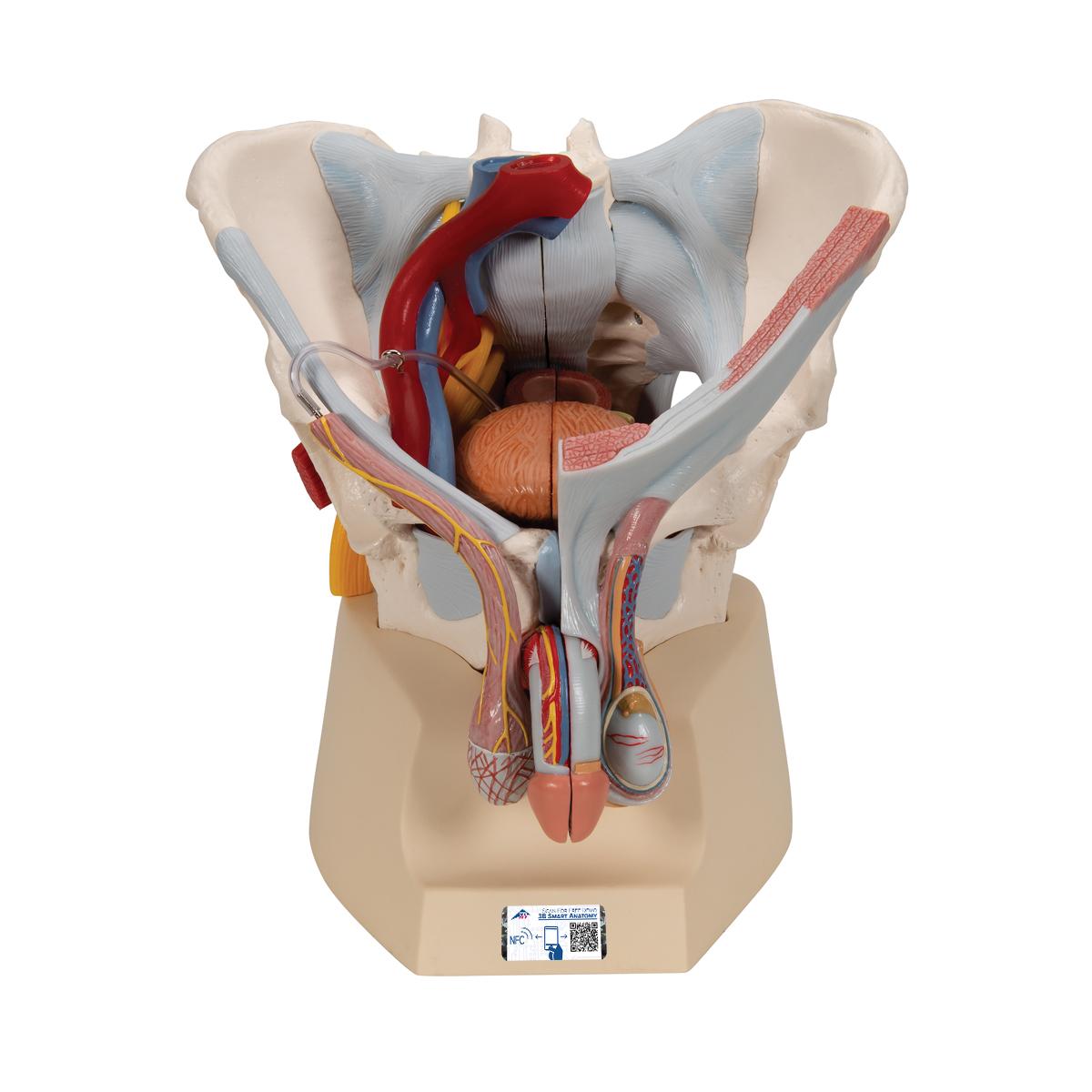
Male Pelvis Skeleton Model with Ligaments, Vessels, Nerves, Pelvic Floor Muscles & Organs, 7 part – 3B Smart Anatomy
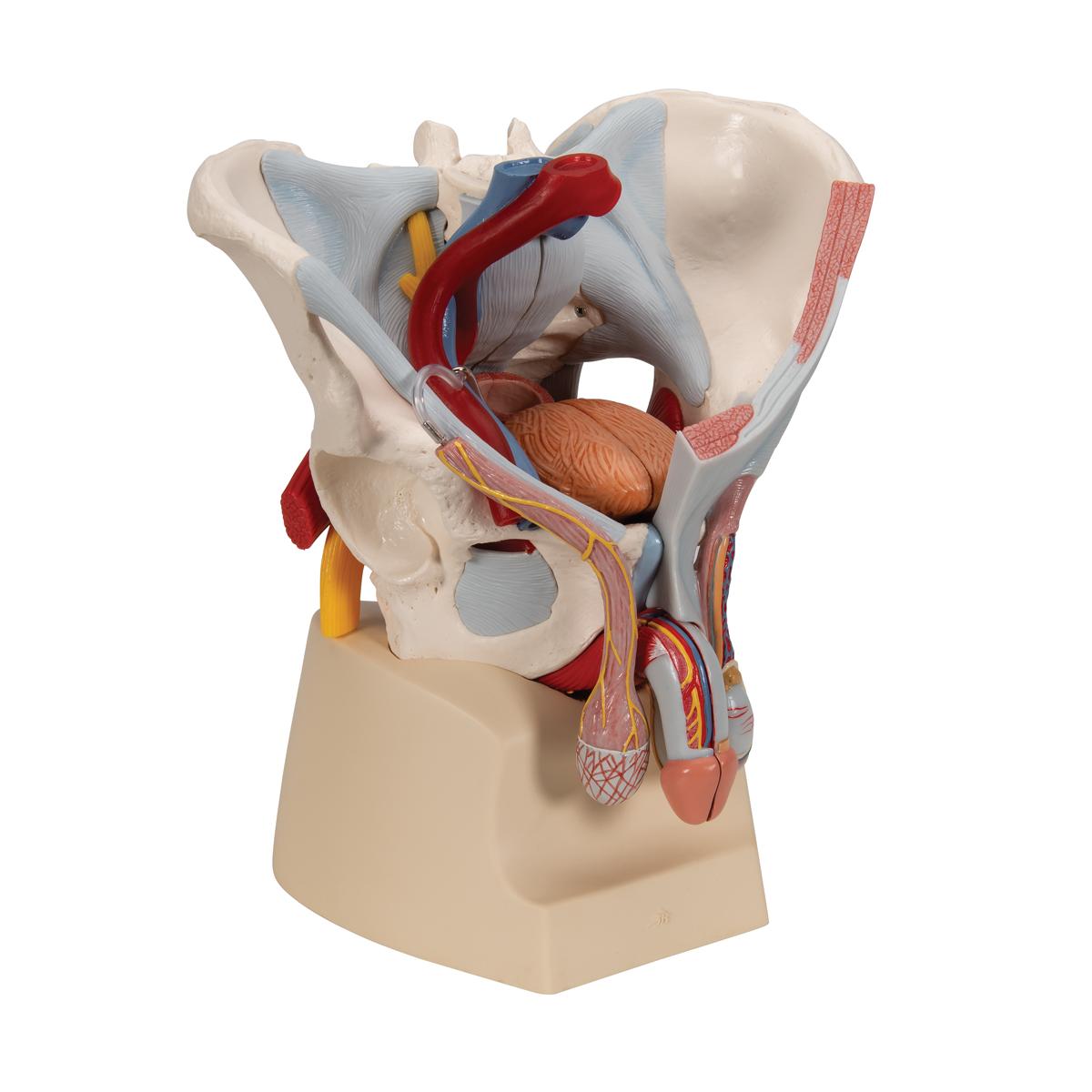
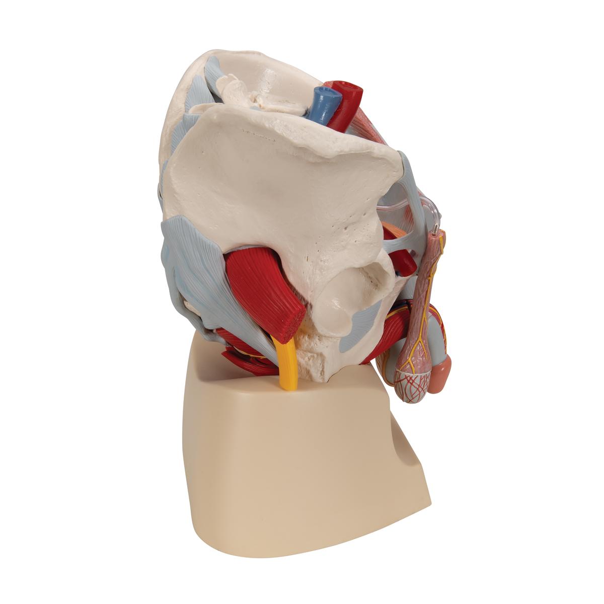
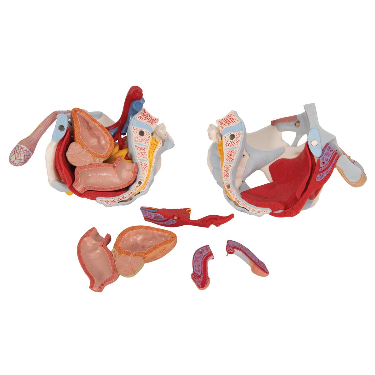
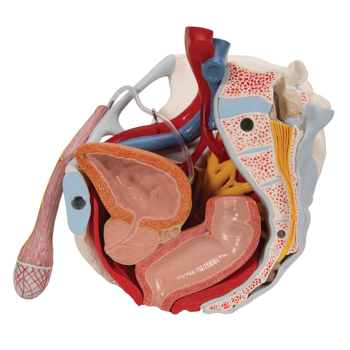
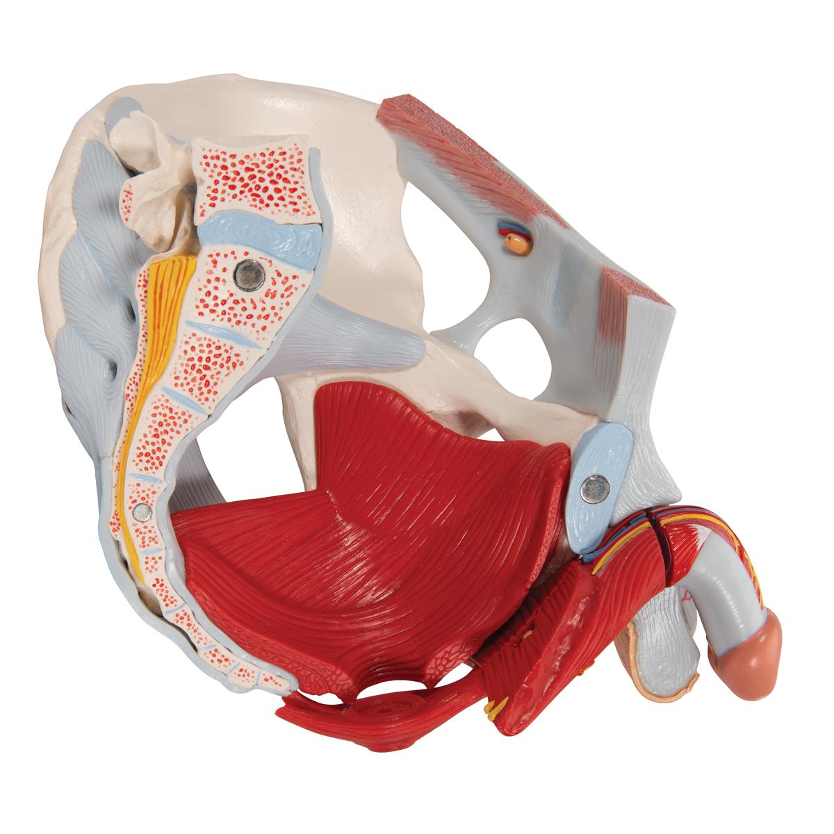
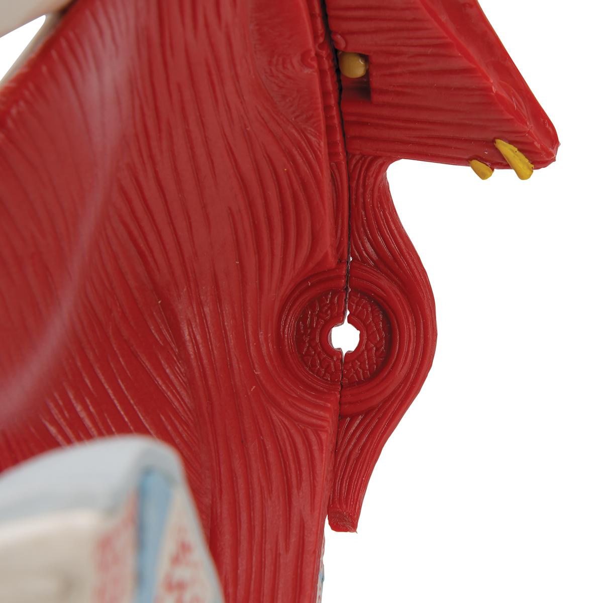
This 7 part model of the male pelvis shows in accurate detail how the bones, ligaments, vessels and nerves as well as the pelvic floor muscles and the external sex organs are connected to each other. It shows the whole pelvis, through which a median section has been placed. The right side of the external anal sphincter, the M. ischiocavernosus, the M. transversus perinei profundus and superficialis and the M. bulbospongiosus can be removed together.
The rectum, bladder, prostate and penis can also be removed, and split into two halves at median level – partially connected. The bone structures are connected with magnets and can therefore be easily taken apart. The skin and the fascia of the penis have been partly removed so that the vessels and nerves are visible. Part of the skin in the area of the scrotum and the spermatic cord has removed too. The testicles and epididymis are also visible. On the left, the spermatic cord has been opened up in layers, and on the right the M. cremaster and fascia spermatica interna have been exposed.
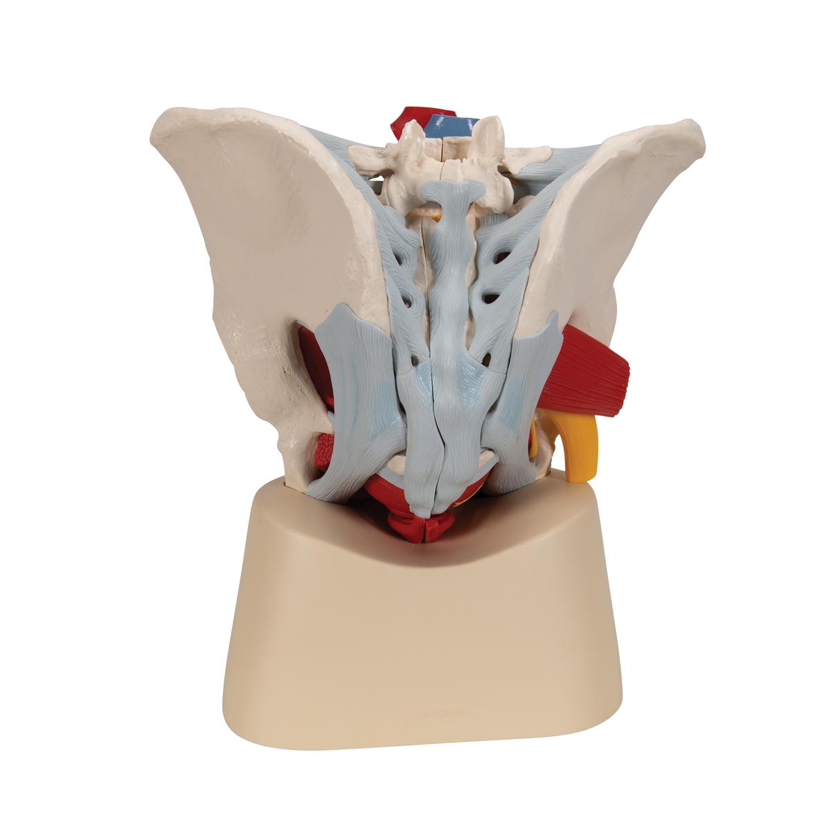
The right half of the pelvis shows sections of the common, external and internal iliac arteries and the common and external iliac veins, demonstrating their positions in relation to each other. The Plexus sacralis, the N. ischiadicus, the N. pudendus, the N. dorsalis penis, the Nn. scrotales anteriores, the Nn. perineales and the Nn. annales inferiores as well as the Ductus deferens are shown. The model shows the following bones and ligaments: both hip bones, pubic symphisis, sacrum and coccyx as well as the fifth lumbar vertebra with intervertebral disc. A median section has been placed through the fifth lumbar vertebra, the sacrum and the coccyx, so that the pelvis can be split into two halves. This means that part of the cauda equina is also visible in the vertebral canal. The left half of the fifth lumbar vertebra can be removed.
The following ligament structures of the pelvis are shown:
- Lig. inguinale
- Lig. sacrotuberale
- Lig. sacrospinale
- Lig. sacroiliaca anteriora
- Lig. iliolumbale
- Lig. longitudinale anterius
- Lig. Supraspinale, Lig. sacroiliacum interosseum
- Lig. sacroiliacum posterius
- Lig. sacrococcygeum laterale
- Lig. sacrococcygeum posterius superficiale et profundum
- Membrana obturatoria
- Lig. lacunare
Every original 3B Scientific anatomy model now includes these additional FREE features:
- Free access to the anatomy course 3B Smart Anatomy, hosted inside the award-winning Complete Anatomy app by 3D4Medical
- The 3B Smart Anatomy course includes 23 digital anatomy lectures, 117 different virtual anatomy models and 39 anatomy quizzes to test your knowledge
- Bonus: FREE warranty upgrade from 3 to 5 years with every product registration
TIP: You will also receive access to a free 3-day trial to all premium features of the Complete Anatomy app when you sign up for your 3B Smart Anatomy course.
To unlock these benefits, simply scan the label located on your model and register online. All 3B Smart Anatomy features are completely free of charge for you. Click here to learn more.